Sunday, May 25, 2025

Unseen Warriors: Cheri Ambrose and the Global Fight Against Male Breast Cancer

By: Lennard M. Goetze, Ed.D

Introduction:
In the vast world of cancer advocacy, few have dared to champion a cause so overlooked, so misunderstood, and so deeply stigmatized. Yet, from the shadows of silence and misdiagnosis emerged a fearless voice—Cheri Ambrose. With unwavering determination and boundless compassion, she has dedicated over a decade to shining a light on a rarely spoken truth: men get breast cancer too. As the founder and driving force behind the Male Breast Cancer Global Alliance, Cheri has transformed personal conviction into a worldwide movement of awareness, support, and lasting change.


Champion of the Voiceless: A Legacy of Bold Compassion

Cheri Ambrose is not a breast cancer survivor herself, but her deep empathy and fierce sense of justice have propelled her to become one of the most important voices in male breast cancer advocacy today. Her story is one of resilience, advocacy, and most of all, humanitarianism. Since 2013, Cheri has committed herself to bringing a voice to those who were silenced by stigma, isolated by fear, or dismissed by systems rooted in bias.

What began as quiet outreach and a desire to fill an information void soon evolved into a full-fledged mission. As she met more men and families grappling with the confusion of a “women’s disease” diagnosis, Cheri saw the devastating effects of societal misunderstanding: late diagnoses, lack of access to clinical trials, emotional alienation, and a complete absence of male representation in public health discussions. For many of these men, Cheri became their first source of validation—and their fiercest advocate.


In 2014, recognizing the urgent need for a centralized resource and community, Cheri founded the Male Breast Cancer Coalition (MBCC), a first-of-its-kind virtual haven for male survivors, caregivers, medical professionals, and advocates. What started as a grassroots initiative quickly gained national attention. The coalition became a beacon for those living in silence, providing not only practical information but also human connection and hope.


By November 2021, the MBCC had grown into a global network, prompting the formation of the Male Breast Cancer Global Alliance (MBCGA). This evolution reflected both the rising number of voices joining the cause and the growing credibility of Cheri’s leadership in the medical and advocacy communities. MBCGA expanded its focus to include not just support, but also scientific collaboration, research advancement, and clinical inclusion. Under Cheri’s guidance, MBCGA now connects survivors, researchers, clinicians, and pharmaceutical leaders from around the world to revolutionize care for men with breast cancer.

Cheri’s humanitarian work doesn’t stop at awareness—it takes tangible form in systemic change. She has been a leading force in persuading pharmaceutical companies to include men in the labeling of breast cancer medications once thought suitable only for women. Through persistence and strategic partnerships, she has opened the door for men to be included in clinical trials, long denied to them due to outdated gender assumptions. Her work not only helps save lives—it rewrites the rules of inclusion in modern medicine.

Advertisement
Mentoring a Movement & Shaping the Next Generation of Advocates
More than an advocate, Cheri is a mentor and mobilizer. She has trained and developed hundreds of survivor representatives—men who now share their stories in hospitals, classrooms, government hearings, and international summits. These men, once voiceless, are now empowered leaders in their own right, thanks to Cheri’s vision and coaching. Their voices form a chorus that breaks through stigma, reshaping public understanding one conversation at a time.

In recognition of her tireless work, Cheri was awarded the Heart of Advocacy honor by the National Consortium of Breast Centers. This prestigious award reflects not just her achievements, but the genuine passion and humanity she brings to every aspect of her mission.

Beyond advocacy, Cheri plays a critical role in the broader cancer awareness movement. As Co-Editor of the New York Cancer Resource Alliance (NYCRA), she contributes to and curates a variety of health and wellness publications including Prevention101.org, NYCRA News, Health Scan News, and First Responders Health Report. Her writing and editorial work amplify her mission, reaching a wide array of professionals, patients, and policy-makers with essential health information and survivor stories.

Cheri is also a graduate of the National Breast Cancer Coalition’s PROJECT LEAD program, and she continues to serve as a key presence at major health summits such as the National Comprehensive Cancer Network Summit, ASCO, SABCS, and the ABC Global Conference in Lisbon. At each of these platforms, she represents not only male breast cancer patients, but all those fighting for recognition and equality in healthcare.

Building a Global Network of Support
Under Cheri Ambrose's leadership, the MBCGA has cultivated a robust network of partnerships with organizations dedicated to providing comprehensive support for men diagnosed with breast cancer. These collaborations aim to address the multifaceted needs of patients, from emotional support to financial assistance.

Financial Assistance through The CARE Project
Recognizing the financial burdens that often accompany a cancer diagnosis, the MBCGA partners with The CARE Project, Inc., a nonprofit organization founded by a breast cancer survivor. The CARE Project's MEN 2 Program offers financial assistance to male breast cancer patients, covering essential expenses such as insurance co-pays, utilities, rent, groceries, fuel, and transportation to and from treatment. This support allows patients to focus on healing without the added stress of financial hardship. 

Educational Resources and Awareness Campaigns
To promote early detection and awareness, the MBCGA has developed multilingual Self-Exam Cards for Men, available in nine languages, including English, Spanish, French, Italian, Dutch, German, Portuguese, Hebrew, and Polish. These cards provide easy-to-follow diagrams and instructions, highlighting warning signs and risk factors specific to men. The initiative is supported by pharmaceutical companies such as Daiichi Sankyo, Inc., Lilly, and Pfizer.

The MBCGA collaborates with leading healthcare institutions to advance research and clinical trials focused on male breast cancer. By bringing together patients, researchers, clinicians, and oncologists, the alliance aims to improve treatment options and outcomes for men diagnosed with the disease. 

In its mission to provide holistic support, the MBCGA partners with organizations such as Imerman Angels, which offers one-on-one support to cancer patients and caregivers through a network of trained Mentor Angels. These mentors provide emotional support and guidance, helping patients navigate their cancer journey. Additionally, the alliance collaborates with Learn Look Locate, an organization dedicated to breast cancer education and support. Together, they work to increase awareness, provide educational resources, and connect patients with support networks. 


Conclusion:

Cheri Ambrose’s legacy is not defined by accolades or titles—it is defined by lives changed, silences broken, and injustices righted. In a world where the voices of the marginalized are often drowned out, she has become a force of amplification. Her compassion is bold. Her fight is personal. Her impact is global.

She has stood up for those who were told they didn’t belong in the breast cancer narrative, and through her, they now have not only a place—but a platform. From living rooms to research labs, from local communities to international health councils, Cheri has made one thing clear: breast cancer doesn’t discriminate, and neither should we.

Cheri Ambrose is more than an advocate. She is a changemaker, a humanitarian, and a beacon of hope for those navigating the darkest moments of their lives. In giving a voice to the voiceless, she has forever changed the story of male breast cancer—for the better.


_________________________________________________________________________________

Sponsor


For more information about the MALE BREAST CANCER GLOBAL ALLIANCE PREDISPOSITION TESTING PROGRAM, contact us at: www.mbcglobalalliance.org or contact our hotline at: 516.522-0777


 --------------------------------------------------------------------------------------------------------------------------

THIS MESSAGE IS BROUGHT TO YOU BY THE MALE BREAST CANCER GLOBAL ALLIANCE

The Male Breast Cancer Global Alliance (MBCGA) is leading the charge in awareness, education, and support for men affected by this disease. This organization has built a worldwide network of survivors, advocates, researchers, and healthcare professionals working to shatter the stigma and silence surrounding male breast cancer. They’ve played a crucial role in pushing for more inclusive research, advancing public health messaging, and ensuring men have access to the resources they need. Through tireless advocacy and collaboration, MBCGA has helped get male breast cancer recognized in global cancer policy and has elevated the voices of countless survivors. Their data-driven campaigns and survivor-led storytelling have reached millions, and their partnership with Bard Diagnostics is all about scaling that impact through accessible genetic testing.

  --------------------------------------------------------------------------------------------------------------------------

RELATED RESOURCES

Under a dedicated partnership with the Male Breast Cancer Global Alliance, BARDDIAGNOSTICS established key programs including: EARLIER DETECTION, PREDISPOSITION GENE TESTING, FIGHT RECURRENCE, ACTIVE SURVEILLANCE AND THE 2ND OPINION SCAN. For the many MEN who seek a significantly improved lifestyle or an upgrade in personalized healthcare, getting better answers about your state of health is paramount and IT ALL STARTS WITH STATE OF THE ART DIAGNOSTICS
. The visionary paradigm that makes up true personalized medical care involves the integration between modalities, a collaboration between specialists and an upgrade in their strategic approach and you'll find it all at BardDiagnostics. 

 --------------------------------------------------------------------------------------------------------------------------

UNCOVERED – Shedding Light on Male Breast Cancer
The first official newsletter from the Male Breast Cancer Global Alliance, launched in proud partnership with AngioMedical Media and the Integrative Cancer Resource Society. Rooted in the belief that education is powerUNCOVERED delivers essential news, scientific updates, and survivor stories to inform and inspire. Each issue is packed with the latest in male breast cancer research, treatment innovations, and advocacy efforts from around the globe. Whether you're a patient, caregiver, or medical professional, UNCOVERED is your trusted source for facts and forward-thinking perspectives. Join us in uncovering the truth—and empowering lives through knowledge. (visit our regularly updated Newsletter)

 --------------------------------------------------------------------------------------------------------------------------

The INTEGRATIVE CANCER RESOURCE SOCIETY is a self-funded (Linkedin Based) independent volunteer group of non-profit foundations/charities, researchers, educators, community leaders and survivors. Under the spirit of collaboration and partnership, we are joined to bring a new level of support to cancer patients, survivors and all those seeking current information about cancer care. We form a unique network of support for one another- while driven to help those who need additional resources, technical updates or empowerment on the road to recovery. ICRS uses the power of the "interweb" to reach a global audience and a network of resources beyond our local borders. We have engaged some of the most impressive minds, perspectives and resources and enjoyed the exchange of vital information that is useful to all. Thanks in part to digital collaboration, these "foreign" connections have always been a part of our cancer crusade, now joining us in what we call "BORDERLESS MEDICINE".

 --------------------------------------------------------------------------------------------------------------------------


In great support of a the global movement of NON-INVASIVE medicine, we proudly introduce the INTEGRATIVE HEALTH RESEARCH CENTER by AngioInnovation Research (501c3). This New York city-based diagnostic imaging validation lab is established to clinically identify the efficacy, performance and supportive claims set by non-invasive therapeutics and diagnostic solutions. Co-founded by Dr. Robert L. Bard, the IHRC offers a non-profit, non-commercialized testing option for health-related innovations who seek an independent and impartial reporting through single-case pilot studies or IRB-Based clinical trials. The IHRC employs credentialed medical professionals and specialists in clinical research whose experience contributes to the unique strategic approach of each test study. (Visit: the AngioInstitute website)

Monday, May 19, 2025

LASER GUIDED VSELs- A Patient's Journey to Restorative Medicine

KINESIOLOGICAL ANALYSIS:  VSELs TREATMENT PROGRESS EXPLAINED

By:  Matthew Einsohn, ND

This might seem like a small thing, but it's actually a big deal. When you're sitting, lifting your leg involves working against gravity, which is already challenging. But when you're lying down, it requires a whole different set of muscles and coordination. It means engaging not just the hip flexors but also the core and stabilizing muscles of the lower back and pelvis. This shows his overall muscle function and coordination have significantly improved.

Without the help of gravity, his core muscles have to work even harder to stabilize and support the movement. This improvement in core strength is great news. Plus, this movement indicates improved flexibility and range of motion, likely due to decreased muscle stiffness and better neuromuscular control.

Being able to perform this movement both sitting and lying down means his nervous system and muscles are working together more effectively. This is crucial for overall mobility and daily activities.

When Dr. Blatman is discussing the origins of pain, which is associated with sheer force and pressure, it appears that stabilization has been achieved. Based on watching the video (above) several times, I would theorize that significant restoration is occurring. It seems he has addressed the root cause of the pain by restoring the integrity of the fascia.

Additionally, while there is a notion that one can make injuries less noticeable, this approach does not seem to align with his method. Instead, if he is indeed focusing on restoring the integrity of the fascia, it suggests that he is addressing the fundamental issue underlying the pain rather than merely masking the symptoms.

Some conditions just this mechanism can help with:

* Muscle weakness and atrophy from post-stroke rehabilitation or muscle degenerative diseases
* Neurological disorders such as MS or Parkinson's
* Recovery from spinal injuries
* Any chronic pain such as Fibromyalgia
* Anyone with joint or mobility problems

Muscle Weakness and Atrophy: Conditions causing muscle weakness or atrophy, such as post-stroke rehabilitation or muscle degenerative diseases, might see improvements in strength and coordination with similar treatments.

Neurological Disorders: Disorders like multiple sclerosis or Parkinson's disease, where muscle control and coordination are affected, could benefit from enhanced muscle function and coordination.

Post-Surgical Rehabilitation: Recovery from surgeries, especially orthopedic or spinal surgeries, could be supported by treatments that improve muscle strength, flexibility, and functional movement.

Chronic Pain Conditions: Conditions like fibromyalgia or chronic back pain that involve muscular and structural issues might see relief through better muscle engagement and reduced stiffness.

Balance and Stability Issues: Treatments that enhance core strength and overall muscle coordination can help with balance and stability, which is crucial for individuals with vertigo or balance disorders.

Joint and Mobility Problems: For conditions like arthritis or other joint issues, improving range of motion and muscle strength can reduce pain and enhance mobility.

Sports Injuries: Athletes recovering from injuries might benefit from improved muscle function and coordination, aiding in a quicker and more effective recovery.


THE BACK STORY: 

VSELs by definition are Very Small Embryonic-Like Stem cells.  They are rare stem cells found in adult tissues that may have potential in regenerative medicine. They are slightly smaller than red blood cells and are located in the inner cell mass of the blastocyst during early development. VSELs are quiescent but can be activated during stress and mobilized into peripheral blood. They have a distinct morphology with a high nuclear/cytoplasmic ratio and undifferentiated chromatin. VSELs also express markers of pluripotent stem cells, such as Oct4, Nanog, and SSEA, and markers of primordial germ cells (PGCs), which suggests they may be related to PGCs. 

Source: https://www.ncbi.nlm.nih.gov/pmc/articles/PMC6461217/


PROCEDURE REPORT: DR. BLATMAN'S TRANSCRIPT ON DR. BARD'S VSELs TREATMENT DAY 1

Dr. Bard and I started the procedure with a deep discussion, making sure we had his wish list and an understanding of what he was specifically wanting and trying to get better. By the time we proceeded, we drew eight tubes of blood, we spun it down, and concentrated his plasma. And then we lasered that plasma and prepared the VSELs out of the solution, activating dormant, very small embryonic like stem cells from his blood -dormant since his birth. And then we prepared them for injection into him. We used eight syringes of these VSELs to inject specific parts of him. There were tendons anchoring his right quadriceps. There were tendons anchoring both of his butt muscles. There were muscles and tendons anchoring the end of his quadriceps that help him control his knee whose weakness brings him knee pain.

I believe he (Dr Bard) was able to tell that when we were doing that needle surgery to place the VSELs so they could facilitate repair of the injured fascia anchors, he could tell that the needle was in the right place. When you've experienced this pain for so long and you can feel what's going on inside your body, you have a way of knowing that I'm in the right spot. And since this procedure can be very uncomfortable, he was able to breathe nitrous oxide which greatly reduces discomfort and anxiety. This made the process a whole lot less uncomfortable for him. And then we followed the needle surgery with an intravenous injection giving him a little more than 40 CCs of the VSELs derived from his blood. These were given intravenously along with an ozone IV at the same time. So the “babies were piggybacked” into the ozonated blood IV after his blood went through ultraviolet light to clean it. As these cells were going into his body, we used a SONG modulated laser to guide these cells to where we want them to get stuck and where we want them to do their work. And so we sent these to his brain, to his cerebellum and balance centers, down his spinal cord, and then into his right thigh.

I'm hoping to see if this technology could reactivate some of the nerves that were damaged from the polio, because when polio damages nerves, it doesn't interrupt or cut them. They're still there, they just don't work. So we're hoping that they're still there enough that we can coax them to work or coax them to health. And we don't know if there's a chance to do that or not, because to the best of my knowledge, this has never been done before (with Post-Polio syndrome). So we are the first ones I would think, to use VSELs in polio for the regeneration of the nervous system and to see if it would work.

When we inject into joints, you can see the cartilage vertical dimension increase. When you inject into rotator cuff tendon, you can subsequently see with ultrasound that the tendon defect filled in. You can also see it on MRI that the defect in the tendon filled in.

We have some patients with Parkinson's in the treatment group from this laser that have shown improvement in 30 minutes. So it can happen pretty quickly. It can also take two or three months before you see a change. You never know.


2024 MEDICAL INNOVATOR: MEET DR. HAL BLATMAN 

Introduction: In the continued search for innovative healers and luminaries in the world of wellness, the Integrative Pain Healers Alliance honors professionals who conduct clinical leadership in their daily practice.  These role models inspire and pave the way for others within their generation to advance their education and expand their capacity in their selected healing arts, or explore other modalities that add to their ability to help their patients.

Our directors and publishers have reviewed the integrative work of Dr. Hal Blatman.  We recognize his holistic & innovative philosophies, his intuitive clinical approach and his expansive vision to engage in what his patients and colleagues would consider “the most sensible and intelligent solutions”.

With dual locations in Cincinnati, Ohio and NYC, Dr. Blatman founded the Blatman Health and Wellness Center- offering a variety of the latest protocols and modalities. His featured health services include: Regenerative Medicine (Stem Cell therapy & PRP), Pain Management and IV Therapy.  Dr. Blatman has also established a successful history working with patients with complex medical disorders by exploring a wider set of diagnostic and therapeutic options and exploratory solutions which includes Metabolic Terrain Medicine for those with cancer. He is also certified in the ReCODE program from Apollo Health for those with dementia and Alzheimer's Disease.


"THE BLATMAN METHOD"

Over the course of more than 30 years in clinical practice Dr. Blatman has developed The Blatman Method – a well published strategy for Understanding and Treating Pain. This method is based on 5 rules:

1. You cannot believe pain comes from where you feel it. Your headache does not come from your head, blood vessels or nerves. Pain in your left arm could be a heart attack, and knee pain does not come from the joint, evidenced by persistent knee pain long after joint replacement surgery.

2. It is not important to distinguish what you think the pain feels like. In fact, your brain cannot tell the difference between sharp, dull, achy, stabbing, numbness, tingling, burning, and more. They all can have the same cause.

3. The only thing you can really believe is what you can touch and feel, and where you are specifically tender, millimeter by millimeter, is where your fascia is kinked or tied in a knot, or where your fascia anchors to hold you together and you have injured and repaired this hundreds of times.

4. If you are examined this way looking for where you are tender from head to toe, the places where you are overall most tender represent the kinks and injuries from your lifetime that generate most of the pain of which you are conscious.

5. No matter how long you have had the pain, and no matter what you have been told is your diagnosis – as quickly as you unkink your fascia and get your body to regrow and strengthen to hold together better on the inside – the pain you had will already be gone.


REGENERATIVE MEDICINE 

Dr. Blatman has been helping people recover and restore worn joints and avoid joint replacement surgery for more than 30 years. His patients have seen great success with regenerative and holistic therapies for pain. Regenerative medicine therapies include Peptide therapy, Platelet-Rich-Plasma (PRP) Therapy, Stem Cell Therapy, exosome treatments, and vSEL Therapy.

Painful and degenerative joints aren’t nearly the only applications of regenerative medicine:

• Tendon repair – Rather than cutting and anchoring injured tendons with surgical repair, regenerative techniques involve reinjuring the tendon with a needle surgery technique and injecting growth factors that facilitate biologic repair of the tendon.

• Ligament repair – Rather than cutting and anchoring injured ligaments with surgical repair, regenerative techniques involve reinjuring the ligament with a needle surgery technique and injecting growth factors that facilitate biologic repair of the ligament

• Joint cartilage restoration – Rather than replace a worn joint via invasive surgery, use the idea that our bodies restore cartilage from daily wear and tear minute by minute, and enhance this process with glucosamine, peptides, PRP, and stem cells.

• Sexual dysfunction treatment – Regenerative medicine techniques may involve injecting growth factors (PRP/stem cells) into intimate areas to regenerate blood vessels and nerves that bring improvement with engorgement and sensation.

• Pelvic floor repair – Growth factor injection and radiofrequency treatments can reduce urinary stress incontinence in women by aiding in repairing and strengthening the pelvic floor and associated muscles.

• Pelvic pain recovery – Regenerative treatments can be used in addition to physical therapy to facilitate repair of fascia that anchors pelvic, gluteal, and thigh muscles. This includes chronic pain generally attributed to the pudendal nerve.

• Heart repair – When heart function has been lost from injuries like ablation and heart attacks, laser-activated and SONG modulated laser-guided vSELS (very small embryonic-like stem cells) can restore heart function and have even gotten people off the heart transplant list.

• Lung repair – Stem cell therapies have been used for more than 10 years to help restore lung tissue and function in people with degenerative lung conditions like COPD and emphysema.

• Neurorehabilitation – Part of our treatment for neurological disorders includes peptides and IV injections of laser activated and laser guided vSELS (very small embryonic like stem cells). We have seen improvements in people with MS, Parkinson’s, and Alzheimer’s.


BIO & BACKGROUND: 

HAL BLATMAN, MD, DAAPM, ABIHM

Dr. Blatman is a nationally recognized specialist in treating myofascial pain. He is credentialed in Pain Management and Board Certified in Occupational and Environmental Medicine and in Integrative Medicine. Dr. Blatman is a Past-President of the American Holistic Medical Association. After receiving his medical degree from the Medical College of Pennsylvania in 1980, Dr. Blatman completed two years of training in orthopedic surgery. He later studied clinical toxicology and ergonomics during his residency in Occupational and Environmental Medicine at the University of Cincinnati Hospital. For several years, Dr. Blatman practiced family medicine while managing a chain of urgent care facilities. During Dr. Blatman’s tenure in general medicine, he recognized standard of care medicine’s difficulty treating and managing chronic and myofascial pain. In his search for answers he studied with the late Janet Travell, MD, then recognized as the world’s leading expert in treating myofascial pain disorders. He now teaches doctors and other providers, and leads a clinical team that understands that pain generally comes from a lifetime of injuries to fascia, and by healing most of it can be made to go away.

For more information, visit Dr. Blatman’s website at: www.blatmanhealthandwellness.com


Sunday, May 18, 2025

A Diagnostic Partnership: The Synergistic Power of Thermography and Ultrasound Imaging


In today’s evolving landscape of diagnostic medicine, the integration of complementary imaging technologies is redefining how clinicians detect, assess, and monitor disease. Two non-invasive modalities—medical-grade thermography and diagnostic ultrasound—have individually proven their worth in clinical settings. Yet, when these tools are combined, their collective diagnostic power offers a more comprehensive understanding of physiological and anatomical conditions, enhancing the accuracy and effectiveness of patient care.

This article highlights the valuable alliance between thermography and ultrasound imaging, illustrating how thermography’s functional, heat-based data guides targeted, structural evaluation through ultrasound. Additionally, we explore how Doppler ultrasound’s capability to assess vascular patterns aligns with thermographic readings, offering a dynamic, multi-dimensional perspective of pathology. Together, these technologies support early detection, monitoring, and integrative care strategies across a range of clinical applications.

Thermography: Capturing the Body’s Thermal Signals
Medical thermography operates by detecting and mapping infrared heat emissions from the body’s surface. Because changes in circulation, inflammation, and nerve activity affect skin temperature, thermography can identify subtle physiological changes that may indicate developing disease, often before structural abnormalities are detectable by other imaging modalities.

Widely known for its role in breast health assessments, thermography is valued for its radiation-free, non-invasive nature and its capacity to detect abnormal heat patterns related to increased vascular activity or inflammation. Beyond breast imaging, thermography is increasingly applied in vascular screening (such as carotid artery assessments), thyroid dysfunction evaluations, and musculoskeletal and nerve-related conditions.


Primary advantages of thermography include:

    * Detection of early physiological changes that precede structural issues.

    * Non-invasive, radiation-free imaging suitable for regular monitoring.

    * Identification of heat patterns associated with abnormal vascularity, inflammation, or nerve irritation.

    * Ability to perform comparative, sequential imaging to monitor physiological trends over time.

Although thermography is not intended to replace anatomical imaging, its sensitivity to functional abnormalities makes it a valuable tool in proactive and complementary diagnostics.


Ultrasound Imaging: Anatomical Insight and Dynamic Evaluation
Diagnostic ultrasound uses high-frequency sound waves to create real-time images of internal tissues and structures. Its versatility allows clinicians to visualize soft tissue anatomy while also assessing movement and blood flow using Doppler technology.

Ultrasound is routinely used for evaluating breast, thyroid, vascular, abdominal, pelvic, and musculoskeletal health, among other applications. Doppler ultrasound adds a critical dimension by enabling clinicians to observe blood flow patterns, vessel integrity, and abnormal vascular activity such as tumor angiogenesis or inflammatory hyperemia.

Notable strengths of ultrasound imaging include:

    * Immediate, real-time visualization of anatomical structures.

    * Functional assessment of blood flow with Doppler imaging.

    * Safe, non-invasive, and adaptable for various anatomical regions.

    * Guidance for needle biopsies and interventional procedures.

Ultrasound’s ability to combine structural detail with dynamic functional data makes it an ideal counterpart to the physiological information captured through thermography.


Thermography as a Guide for Targeted Ultrasound Evaluation
The value of pairing thermography with ultrasound lies in how thermal imaging can direct more precise, focused ultrasound assessments. Thermography highlights areas of abnormal heat distribution, which often correspond to regions of increased blood flow, inflammation, or nerve activity beneath the skin. These thermal findings can then inform where ultrasound should be applied, ensuring that subclinical or early-stage concerns are investigated further.

In clinical scenarios such as:

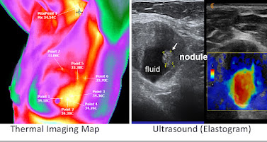
    * Breast health assessments: A localized heat anomaly on thermography may indicate increased vascular activity. Focused ultrasound imaging, supplemented with Doppler studies, can then examine the area for masses or abnormal vascularity.

    Carotid artery evaluation: Thermal asymmetries along the neck may signal vascular irregularities or inflammation. Doppler ultrasound can assess blood flow velocities and vessel patency at these flagged locations.

    Thyroid screening: Thermal patterns suggestive of hyperactivity or inflammation can prompt targeted ultrasound evaluation to visualize nodules, cysts, or vascular changes.

This coordinated approach improves diagnostic accuracy by combining thermography’s sensitivity to functional changes with ultrasound’s structural and functional imaging capabilities.


Aligning Vascular Data: Doppler Ultrasound Meets Thermography
Doppler ultrasound’s role in measuring blood flow dynamics aligns naturally with the vascular insights offered by thermography. Both modalities offer distinct, yet complementary, information about circulation and tissue metabolism.

For example:

    * Tumor-related angiogenesis often results in increased local blood flow, creating areas of hyperthermia on thermography and detectable vascular proliferation with Doppler ultrasound.

    * Inflammatory processes generate heat due to increased blood flow and metabolic activity. Thermography can map these hotspots, while Doppler ultrasound confirms hyperemia and assesses the involved vessels.

    * Peripheral vascular disease may appear as cooler regions on thermography due to impaired perfusion. Doppler ultrasound can then quantify blood flow reduction or identify vascular obstructions.

    * The alignment of these two data sets provides clinicians with a thorough understanding of both functional vascular health and anatomical vessel status.


Broadened Applications: Established and Investigational Uses
While thermography holds FDA clearance as an adjunct for breast health evaluation, practitioners have extended its use into several investigational areas where early physiological changes are clinically relevant.

Examples include:

    * Neuropathy and pain syndromes: Thermography can detect altered heat patterns associated with nerve irritation or dysfunction in the back, feet, and pelvic areas.

    * Inflammatory arthritis and rheumatic conditions: Localized heat increases can identify active inflammation sites.

    * Post-surgical surveillance: Thermography may help track vascular integrity and inflammation following operative procedures.

    * Sports injuries and overuse syndromes: Subtle thermal changes can signal early-stage tissue stress or inflammation, while ultrasound evaluates tissue integrity and guides treatment.

In each scenario, ultrasound enhances the diagnostic process by confirming structural pathology or assessing blood flow in areas identified by thermography, allowing for more precise intervention planning.


Illustrative Example: Integrating Imaging for Breast Health
Consider a clinical case in which thermography reveals a localized area of elevated heat in the upper outer quadrant of the left breast. This finding prompts a targeted ultrasound examination, which identifies a small, hypoechoic mass with notable vascularity on Doppler imaging. The combined results offer a compelling clinical rationale for further investigation, such as biopsy or additional imaging.

In situations where breast tissue density limits mammographic accuracy, thermography and ultrasound together improve the likelihood of early detection, providing valuable information in risk assessment and management planning.


Conclusion: A Forward-Thinking Diagnostic Model
The combined use of medical thermography and ultrasound imaging represents a progressive, integrative model in diagnostic medicine. By coupling physiological heat mapping with real-time anatomical and vascular imaging, clinicians can detect early, often subclinical changes that may signal the onset of disease. This dual-modality approach offers a safer, non-invasive, and highly informative option for proactive health monitoring and early intervention.

As diagnostic medicine continues to evolve, the partnership between thermography and ultrasound promises to play an increasingly significant role, particularly in breast health, vascular screening, thyroid evaluation, and investigational applications in pain, inflammation, and sports medicine. Future research and clinical protocols will further refine and expand these applications, advancing the goal of truly personalized, precision-based care.


RELATED ARTICLES


ADVANCED THERMOLOGY FOR THYROID AND CERVICAL DIAGNOSTIC EVALUATION

THE THYROID GLAND, situated anteriorly in the lower neck, is uniquely accessible due to its superficial anatomical position and rich vascular network. This makes it an ideal candidate for assessment through high-resolution infrared thermographic imaging. Thermology offers a non-invasive, radiation-free adjunctive modality that complements traditional diagnostic methods in evaluating thyroid function and pathology. By detecting subtle temperature variations on the skin surface above the thyroid and surrounding cervical tissues, infrared thermography reflects the gland’s underlying metabolic activity. This technique is especially valuable in the early detection and functional assessment of thyroid disorders, including Hashimoto’s thyroiditis, Graves’ disease, nodular goiter, and thyroid malignancies.


THE ROLE OF NEOANGIOGENESIS IN BREAST CANCER DETECTION

One of the core principles of medical thermography is detecting physiologic changes associated with cancer. Neoangiogenesis—the formation of new blood vessels—is critical for tumor growth. Without it, cancerous cells would be unable to obtain the nutrients they need to proliferate. The abnormal structure of these new blood vessels was not well understood until the late 1980s, when studies revealed their unique porous nature.

Unlike normal blood vessels, which have a structured, three-layered composition, neoangiogenic vessels are disorganized and resemble a natural sponge, with an erratic, unregulated flow. Lacking smooth muscle and autonomic nervous system regulation, these vessels exhibit excessive and uncontrolled blood flow. As a result, areas of neoangiogenesis appear as "hot spots" in thermal imaging. Because breast tissue is located in the outer shell of the body, where body heat is regulated to maintain a stable core body temperature, these hot patterns can indicate malignancy, setting medical thermology apart as a functional imaging modality distinct from structural imaging techniques like mammography, ultrasound and MRI.


HEAD AND NECK: QUANTITATIVE THERMAL MEASUREMENT & A MEDICAL COMPLETEMENT TO THE ULTRASOUND

The patient is a 65-year-old military veteran who sustained a fall, leading to a concussion that required a three-day hospital observation period. Upon discharge, he reported persistent shoulder pain. Ultrasound imaging of the shoulder revealed an inflamed biceps tendon, which was appropriately treated with physical therapy or targeted injection—avoiding unnecessary rotator cuff surgery, which had initially been planned. Given the patient’s recent concussion, ultrasound assessment was extended to the optic nerve, which was imaged within minutes and confirmed as normal, ruling out increased intracranial pressure.

Optic nerve swelling is also another a critical indicator of elevated intracranial pressure and is widely used in emergency medicine for concussion assessment. While hospital concussion screenings may clear a patient, they do not eliminate the risk of delayed-onset intracranial bleeding. Subdural hematomas, for example, can develop over days, weeks, or even months post-injury, gradually increasing intracranial pressure. Early detection using noninvasive imaging techniques is therefore essential and timely.


Seen, Heard, Empowered: What My Visit to Dr. Bard Taught Me About Midlife Health

[⚠️ Suspicious Content]

Introduction

The Women’s Health Collaborative MIDLIFE Health Group is proud to present this powerful firsthand narrative by Sabine Gee, a dedicated Midlife Health & Menopause Coach and passionate advocate for women’s wellness. For years, Sabine has guided countless women over 45 through the complexities of perimenopause, seeking out forward-thinking tools and progressive care strategies that go beyond conventional medicine. Yet it was her long-awaited meeting with Dr. Robert Bard in New York City that would profoundly reshape her clinical perspective.

A pioneer in patient-centered diagnostics and the visionary behind the groundbreaking MenoScan, Dr. Bard offers a refreshingly transparent, hands-on approach to imaging and risk assessment for midlife women. Drawn by a shared commitment to addressing the overlooked and often dismissed symptoms of perimenopause, Sabine sought to experience firsthand how advanced imaging could reveal crucial insights often missed in traditional screenings.

This encounter not only deepened Sabine’s understanding of integrated midlife care but forged a meaningful alliance between two advocates committed to empowering women through proactive, personalized diagnostics. Together, their collaborative mission champions a future where women are seen, heard, and equipped with the knowledge to reclaim agency over their health journey.


"A Personal Journey Into Advanced Midlife Diagnostics
 As a Midlife Health & Menopause Coach, I’m always on the lookout for tools, research, and experts that help women over 45 reclaim their energy, confidence, and health. But sometimes, an experience leaves such a strong impression that it shifts your perspective entirely. That’s exactly what happened during my visit with Dr. Robert Bard in New York City.

Dr. Bard is not your typical radiologist. He is a pioneer in patient-centered diagnostics and the visionary behind the MenoScan—a groundbreaking set of advanced imaging protocols designed to identify hidden risk factors in midlife women. What sets him apart isn’t just the technology he uses, but how he uses it: with the patient, not behind the scenes.

Why I Went
As someone navigating midlife myself, I was curious about the real impact imaging could have on health outcomes—not just for me, but for my clients. I wanted to see what the MenoScan could reveal about my body and how it might complement traditional blood tests.

I was greeted warmly and shown around the office by Dr. Bard himself—a rare experience in today’s medical world. He listened attentively as I shared my health history and specific concerns, then tailored the session to fit my needs.

The Front-Row Experience
What followed was nothing short of remarkable. We began with thermal imaging of my hands and feet—checking for signs of inflammation and arthritic changes. He also performed thermal imaging of my neck and my eye—the latter to assess blood flow to the brain, which is a crucial indicator of brain health. From there, we transitioned to a scan of my neck and carotid artery. With each scan, Dr. Bard explained what we were seeing, in real time.

The most powerful moment? When he examined my thyroid and carotid artery while I sat beside him. The screen was visible to both of us. He explained the health of my thyroid, showed me the blood flow, and answered my questions with care. I felt part of the process. Seen. Heard. Empowered.

Why Imaging Matters
So many midlife women struggle with symptoms they don’t understand: fatigue, weight gain, anxiety, hot flashes. And too often, they’re told it’s all "just aging." But that’s not good enough.

Thyroid health, for example, often goes unchecked beyond a standard blood panel. Yet imaging can detect tissue changes or inflammation well before bloodwork reveals any issues. In fact, Dr. Bard explained that non-invasive tools like elastography and Doppler imaging can even help predict malignancy or rule it out, guiding early intervention.

And when it comes to cardiovascular health, many women are shocked to learn that heart disease is the #1 cause of death post-menopause. While I had recommended calcium scans in the past, I was amazed to learn that Dr. Bard's ultrasound-based scan of the carotid artery can detect early vascular changes even before calcium scans show plaque. This is crucial information for women deciding whether HRT is right for them.

It’s important to note that these are just a few of the many tools included in the comprehensive MenoScan protocol. To explore the full range of what the MenoScan offers, visit: www.menoscan.org


A Diagnostic Revolution
Dr. Bard is revolutionizing how we approach diagnostics. In most U.S. clinics, a technician does the scan, a radiologist reads it later, and the results are then relayed to the patient via yet another doctor—a sharp contrast to what I’m used to in Europe, where each doctor typically performs and interprets their own imaging.

Dr. Bard defies this fragmented model. He holds the probe himself, interprets the scans in real time, and makes the patient an informed partner in the process.

He’s also not afraid of potential litigation or protocols that discourage real-time patient dialogue. Instead, he believes in transparency, empowerment, and human connection—qualities that are sadly rare in modern medicine.

As Dr. Bard says:
"We don't just look for disease. We look for patterns, for early changes, for what might become disease. That's where prevention begins.”


The Future of Midlife Health
The MenoScan is more than a test—it’s a foundation for informed decision-making. Combined with bloodwork and a skilled practitioner, it gives women a full picture of what’s really happening inside their bodies. It highlights the multi-factorial nature of menopause: how hormones, cardiovascular function, thyroid health, inflammation, and even mental well-being are interconnected.

Too often, women are prescribed antidepressants during perimenopause when the real issue is hormonal fluctuation. Too often, they’re told “nothing is wrong,” or that symptoms are "just part of aging."

But as I always say: "The ones with symptoms are the lucky ones.”

Because at least their bodies are speaking up, prompting women to look for solutions, so they can take charge of their health and a healthy, active & joyful next chapter. Let’s stop brushing off women’s symptoms. Let’s start offering them real answers, powerful tools, and a seat at the table when it comes to their own health. Thanks to Dr. Bard, I left not only reassured but equipped—with knowledge, images, and insight I could use to take the next step in my health journey. Every woman deserves this.


___________________________________________________________________________________


Epilogue: Building Bridges for Women’s Health Advancement
By: Dr. Barbara Bartlik & Marilyn Abrahamson

In today’s complex healthcare landscape, meaningful change rarely happens in isolation. True progress is born from collaboration—when medical pioneers, health coaches, patient advocates, and wellness professionals join forces in pursuit of a common goal. This narrative reflects the growing importance of building bridges between clinical experts like Dr. Robert Bard and passionate non-medical professionals like Sabine Gee, whose firsthand insights into the lived experiences of midlife women enrich the dialogue around care.

Click to see the full results of this event

Forging these alliances is essential in the ongoing crusade for better education, advocacy, and diagnostic innovation. Women navigating perimenopause and beyond deserve a healthcare model where expertise is shared, silos are dismantled, and new ideas flourish. By networking across disciplines, we foster environments where groundbreaking technologies like the MenoScan reach those who need them most—and where women’s health concerns are met with real solutions, not dismissive platitudes.

The Women’s Health Collaborative MIDLIFE Health Group stands as a testament to this mission. Together, we amplify each other’s work, validate women’s experiences, and accelerate the movement toward comprehensive, patient-centered care. The future of midlife health belongs to those who are bold enough to build it together.


Thursday, May 15, 2025

THYROID HEALTH: WHY ULTRASOUND MATTERS

3-25-2025   By: Dr. Leslie Valle-Montoya

Have you ever had your thyroid checked? If not, now is the time! While standard thyroid function tests measure hormone levels in the blood to detect disorders like hypothyroidism (underactive thyroid) and hyperthyroidism (overactive thyroid), they don’t tell the whole story. That’s where modern ultrasound technology steps in.

Unlike blood tests, which focus on hormone production, ultrasound provides a real-time, detailed view of the thyroid gland itself. This imaging technique uses sound waves to assess the gland’s size, shape, and structure—offering insights that hormone tests simply can’t provide.

Advanced ultrasound methods, such as 3D Doppler Blood Flow and Elastography, enhance diagnostic accuracy by measuring blood flow and tissue stiffness. These features help differentiate between benign and potentially malignant nodules, offering a clearer picture of thyroid health. By identifying whether nodules are solid or fluid-filled, ultrasound aids in assessing cancer risk and guiding further evaluation.

If you've never had a thyroid ultrasound, consider it an essential step in understanding your thyroid health beyond just hormone levels. A complete evaluation means looking at both function and structure—because when it comes to your health, knowledge is power.


Prologue:

LEARNING FROM THE ULTRASOUND GURU - (Day One)

Since COVID, I’ve connected with countless health professionals over Zoom. Even today, virtual meetings remain the norm for work engagements, often replacing in-person visits and hands-on collaborations.

But today, we broke that mold. I had the privilege of finally meeting Dr. Robert Bard in person—a true pioneer in diagnostic imaging! After five years of remote referrals, I traveled from Santa Barbara, CA, to New York City for a firsthand look at his cutting-edge arsenal of medical technology. More than just a tour, I also received an intensive crash course in ultrasound operation. Dr. Bard had previously mailed me a hospital-grade Terason T-3200 for my practice, and now, under his guidance, I was able to put it to use.

This marks the beginning of our TelemedScan remote overreading collaboration, and learning from "The Cancer Detective" himself was an incredible honor—an opportunity few get to experience from the front row.

"The best way to learn ultrasound is to be a patient," Dr. Bard explained. "Its real-time scanning ability lets you see as you scan—the screen tells all, right on the spot!"

With that in mind, we decided to scan my thyroid, a first for me. Given our collaboration on a perimenopause project, I knew it was time to practice what I’m about to preach. Having access to one of the best thyroid imaging experts in the field was an unexpected luxury—one I wasn’t about to pass up. During the session, I was introduced to the concept of sonology—where the radiologist directly controls the probe, eliminating the middle step of a technician. This hands-on approach allows for immediate interpretation, deeper exploration, and a more interactive learning experience between doctor and patient.


TO BE CONTINUED ON MENONEWS:
Common History of Patients Who Benefit from Imaging


ABOUT THE AUTHOR

LESLIE VALLE-MONTOYA, MD is the Founder of Biomed Life and the Santa Barbara Longevity Center. After medical school, Dr. Valle focused on managing chronic disease starting with its links to poor nutrition and then introducing them into the world of energy frequencies.  She explores and includes non-invasive modalities such as: frequency therapies (including biofeedback), PEMF, proper detoxification, nutritional guidance and binaural beats as needed. - visit: www.biomedlifesb.com


The Essentials of Thyroid Imaging (part 2)

By: Dr. Robert Bard

When imaging detects a suspicious area, it can guide selective biopsies to obtain small tissue samples for further laboratory analysis (pathology). Combining imaging with pathology provides the most accurate information about the size, location, and aggressiveness of any issue detected.  In the case of thyroid imaging, use of an ultrasound uses sound waves to create images of the thyroid gland in the neck. It is often used for this organ because it is a non-invasive procedure that does not use ionizing radiation. It’s commonly used to evaluate lumps or nodules detected during a physical exam or other imaging tests and requires minimal preparation.

 Thyroid nodule (tumor) - doppler ultrasound

The ultrasound helps doctors visualize the thyroid's size, shape, and potential abnormalities like nodules, cysts, or inflammation, aiding in diagnosis and treatment planning. It’s typically performed when physical exams show:

 - A growth on the thyroid, known as a thyroid nodule (image - R).

- An enlarged or irregular thyroid (goiter).

- Abnormal lymph nodes near the thyroid.


THE ROLE OF THE THYROID GLAND: The thyroid is a butterfly-shaped organ in the endocrine system that regulates hormones influencing various bodily functions. These hormones affect both physical and mental performance, so unexplained symptoms may indicate a thyroid issue.

HYPERTHYROIDISM AND THYROID DISORDERS: Thyroid disorders can stem from various causes and affect the balance of hormones in the body. A range of treatments is available for managing thyroid issues, from medication to non-invasive procedures.


Part3:

THYROID CANCER:
Thyroid tumors are common but can be hard to diagnose by touch alone. Sonography is the primary method for detecting and monitoring suspicious nodules. Areas with calcium deposits are especially concerning and often require biopsy under ultrasound guidance. Parathyroid masses, which can cause significant health issues like bone loss in women or kidney stones in men, may also be detected. Ultrasound can also identify cancer spread to nearby lymph nodes, which can be biopsied with imaging guidance.


THYROID CANCER FAQ's

Q: Why is a sonogram important if I visit my doctor annually?
Blood tests often miss thyroid tumors, which can be difficult to detect through physical examination, especially if they are deep within the gland.

Q: What happens if a tumor is found?
Many benign conditions, like cysts, can be differentiated from cancer without a biopsy. Ultrasound can assess calcium content, which may indicate cancer, requiring further tests.

Q: Does every suspicious area require a biopsy?
Ultrasound screening and other tests can reduce unnecessary biopsies. Small suspicious areas are typically monitored for growth every 3-6 months before considering a biopsy.

Q: How far can thyroid cancer spread?
Most thyroid cancers grow slowly and spread locally. Metastasis is rare and can be evaluated using MRI scans.

Q: Can ultrasound detect parathyroid tumors that cause osteoporosis?
Parathyroid adenomas are typically benign but can lead to significant physical effects due to hormonal imbalances. These tumors can often be diagnosed with 3-D Power Doppler Histogram .

In summary, ultrasound imaging plays a crucial role in evaluating thyroid health by providing a safe, non-invasive, and highly detailed view of the gland. When combined with pathology, it enhances diagnostic accuracy, guiding appropriate treatment decisions. This synergy between imaging and laboratory analysis ensures a comprehensive approach to identifying and managing thyroid conditions effectively.


ABOUT THE AUTHOR-

ROBERT L. BARD, MD, PC, DABR, FASLMS - Advanced Imaging & Diagnostic Specialist

Having paved the way for the study of various cancers both clinically and academically, Dr. Robert Bard co-founded the 9/11 CancerScan program to bring additional diagnostic support to all first responders from Ground Zero. His main practice in midtown, NYC (Bard Diagnostic Imaging- www.CancerScan.com) uses the latest in digital Imaging technology has been also used to help guide biopsies and in many cases, even replicate much of the same reports of a clinical invasive biopsy. Imaging solutions such as high-powered Sonograms, Spectral Doppler, sonofluoroscopy, 3D/4D Image Reconstruction and the Spectral Doppler are safe, noninvasive, and does not use ionizing radiation. It is used as a complement to find anomalies and help diagnose the causes of pain, swelling and infection in the body’s internal organs while allowing the diagnostician the ability to zoom and ‘travel’ deep into the body for maximum exploration.

......................................................................................................................................................................


A VOICE FROM THE COMMUNITY

Kudos to Dr. Bard for his groundbreaking work with MenoScan, a diagnostic innovation that is transforming how menopause is understood, diagnosed, and managed. By addressing the intricate interplay of hormonal balance, bone density, cardiovascular function, neurological health, and overall metabolic stability, MenoScan empowers both patients and physicians to make informed, personalized decisions around prevention and care.

In collaboration with Dr. Angela Mazza—a triple board-certified endocrinologist and founder of the Metabolic Center for Wellness—Dr. Bard has assembled a team of leading medical experts dedicated to a comprehensive, individualized approach to women’s health during midlife and beyond. Dr. Mazza’s work highlights the importance of personalized thyroid care, particularly during the menopausal transition. As the accompanying graphic illustrates so clearly, menopause impacts all women. Understanding it fully is essential to improving care and outcomes.

- ROBERTA MORRISMenopause Educator l Neuro-Nutrition Specialist


CH10: READING BETWEEN THE ECHOES

THE EYE WITHIN UNLOCKING THE HIDDEN LANGUAGE OF MEDICAL IMAGING By: Lennard M. Goetze, Ed.D   In an age when medical imaging technologie...